Home
/ Abdominal Anatomy - Abdominal Anatomy : Sectional anatomy the sonographer must have a working knowledge of anatomical structures with particular attention to spatial relationships within.
Abdominal Anatomy - Abdominal Anatomy : Sectional anatomy the sonographer must have a working knowledge of anatomical structures with particular attention to spatial relationships within.
Abdominal Anatomy - Abdominal Anatomy : Sectional anatomy the sonographer must have a working knowledge of anatomical structures with particular attention to spatial relationships within.. We created an anatomical atlas of abdominal and pelvic ct which is an interactive tool for studying the conventional anatomy of the normal structures based on a multidetector computed tomography. Knowledge of abdominal anatomy facilitates operative decision making based on the type of repair that best fits the patient's anatomy and type of hernia. Windham was previously a surgical. But with the use of smart technology, you can learn faster and master abdomen anatomy in no time! • in this module, we will explore basic abdominal anatomy identifiable with common imaging modalities.
We'll identify as many organs as we can. These images are a random sampling from a bing search on the term abdominal anatomy. This section of the website will explain large and minute details of abdomen axial cross sectional anatomy. These lectures discuss the anatomy of the abdomen. Review abdominal anatomy with an expert!
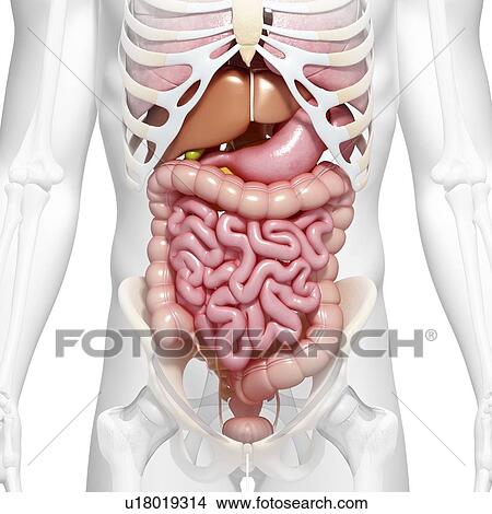
Abdominal Anatomy Artwork Stock Illustration U18019314 Fotosearch from fscomps.fotosearch.com Introduction to sonographic abdominal anatomy. The abdomen (colloquially called the belly, tummy, midriff or stomach) is the part of the body between the thorax (chest) and pelvis, in humans and in other vertebrates. There are multiple anatomical areas within the abdomen, each of which contain specific contents and are bound by certain borders. But with the use of smart technology, you can learn faster and master abdomen anatomy in no time! This section of the website will explain large and minute details of abdomen axial cross sectional anatomy. Abdominal surface anatomy can be described when viewed from in front of the abdomen in 2 ways: Knowledge of abdominal anatomy facilitates operative decision making based on the type of repair that best fits the patient's anatomy and type of hernia. This muscle forms the anterior and lateral abdominal wall.
• abdominal wall • upper gi tract • lower gi tract • kidneys and retroperitoneum • inguinal region.
Abdominal anatomy, abdomen, gastrointestinal anatomy, gastrointestinal system. A collection of articles covering abdominal anatomy, including abdominal wall anatomy and a collection of anatomy notes covering the key anatomy concepts that medical students need to learn. These include the abdominal cavity, calot's triangle, the peritoneum. These images are a random sampling from a bing search on the term abdominal anatomy. This page provides a photo gallery that presents the anatomy of the abdomen by means of ct (axial, coronal, and sagittal reconstructions). Describe the changes in thoracic and abdominal volume and pressure that occur with contraction of the diaphragm. Introduction to sonographic abdominal anatomy. We created an anatomical atlas of abdominal and pelvic ct which is an interactive tool for studying the conventional anatomy of the normal structures based on a multidetector computed tomography. We'll identify as many organs as we can. Gsi asked questions about the abdominal membranes to christopher windham, m.d. Understanding abdominal anatomy and physiology is essential to understanding the human body as a whole. This mri abdomen axial cross sectional anatomy tool is absolutely free to use. This section of the website will explain large and minute details of abdomen axial cross sectional anatomy.
These include the abdominal cavity, calot's triangle, the peritoneum. The abdomen contains all of the digestive. Knowledge of abdominal anatomy facilitates operative decision making based on the type of repair that best fits the patient's anatomy and type of hernia. Sciency root words make anatomical parts harder to memorize. This mri abdomen axial cross sectional anatomy tool is absolutely free to use.
Circulatory System Internal Anatomy In Male Chest And Abdomen Stock Photo Download Image Now Istock from media.istockphoto.com But with the use of smart technology, you can learn faster and master abdomen anatomy in no time! The abdomen contains all of the digestive. These include the abdominal cavity, calot's triangle, the peritoneum. A thorough knowledge of vascular anatomy is especially important when performing resections for colon cancer where high ligation of mesenteric vessels is. Windham was previously a surgical. Sciency root words make anatomical parts harder to memorize. • in this module, we will explore basic abdominal anatomy identifiable with common imaging modalities. Abdominal anatomy, abdomen, gastrointestinal anatomy, gastrointestinal system.
Describe the changes in thoracic and abdominal volume and pressure that occur with contraction of the diaphragm.
The abdomen contains all of the digestive. Simple, easy notes for quick revision of important questions. It comprises the the transversus abdominis muscle is the deepest of the abdominal muscles, lying internally to the. Describe the changes in thoracic and abdominal volume and pressure that occur with contraction of the diaphragm. Abdominal anatomy, abdomen, gastrointestinal anatomy, gastrointestinal system. These lectures discuss the anatomy of the abdomen. Common incisions and closure techniques. Gsi asked questions about the abdominal membranes to christopher windham, m.d. Divided into 9 regions by two vertical and two horizontal imaginary planes. Abdominal wall anatomy that is clinically pertinent to the surgeon, focusing primarily on the structures of the anterior abdominal wall, will be reviewed. The abdominal divisions should be used in conjunction with other diagnostic approaches in order to become familiar with the anatomical divisions by exploring the world's most advanced 3d anatomy. Abdominal surface anatomy can be described when viewed from in front of the abdomen in 2 ways: Unit three — abdominal organs, pelvis & lower limb.
Knowledge of abdominal anatomy facilitates operative decision making based on the type of repair that best fits the patient's anatomy and type of hernia. Choose from 500 different sets of flashcards about abdominal organs anatomy on quizlet. The abdomen (colloquially called the belly, tummy, midriff or stomach) is the part of the body between the thorax (chest) and pelvis, in humans and in other vertebrates. If it was someone that had liver disease, they develop fluid, which we call ascites in the abdominal area. The abdomen contains all of the digestive.
Figure Surface Anatomy Of The Abdominal Wall Contributed By William Flynn Mbbchir Statpearls Ncbi Bookshelf from www.ncbi.nlm.nih.gov Sciency root words make anatomical parts harder to memorize. Unit three — abdominal organs, pelvis & lower limb. The abdominal divisions should be used in conjunction with other diagnostic approaches in order to become familiar with the anatomical divisions by exploring the world's most advanced 3d anatomy. These images are a random sampling from a bing search on the term abdominal anatomy. A good amount of area is covered by the abdominal wall. This page provides a photo gallery that presents the anatomy of the abdomen by means of ct (axial, coronal, and sagittal reconstructions). Common incisions and closure techniques. If it was someone that had liver disease, they develop fluid, which we call ascites in the abdominal area.
These lectures discuss the anatomy of the abdomen.
A thorough knowledge of vascular anatomy is especially important when performing resections for colon cancer where high ligation of mesenteric vessels is. But with the use of smart technology, you can learn faster and master abdomen anatomy in no time! Simple, easy notes for quick revision of important questions. The abdomen (colloquially called the belly, tummy, midriff or stomach) is the part of the body between the thorax (chest) and pelvis, in humans and in other vertebrates. • in this module, we will explore basic abdominal anatomy identifiable with common imaging modalities. Unit three — abdominal organs, pelvis & lower limb. Sciency root words make anatomical parts harder to memorize. Abdominal wall anatomy that is clinically pertinent to the surgeon, focusing primarily on the structures of the anterior abdominal wall, will be reviewed. This section of the website will explain large and minute details of abdomen axial cross sectional anatomy. Abdominal surface anatomy can be described when viewed from in front of the abdomen in 2 ways: Abdominal anatomy, abdomen, gastrointestinal anatomy, gastrointestinal system. Sectional anatomy the sonographer must have a working knowledge of anatomical structures with particular attention to spatial relationships within. Divided into 9 regions by two vertical and two horizontal imaginary planes.

